


Oxidative stress: Specialized markers provide more clues
Dicken Weatherby, N.D. and Beth Ellen DiLuglio, MS, RDN, LDN
Once blood chemistry biomarkers have been evaluated, more specific biomarkers associated with oxidative stress can be utilized. A comprehensive approach would include looking at the following [1]
The ODX Oxidative Stress Series
- Oxidative Stress part 1 - And You Thought You Were Stressed!
- Oxidative Stress part 2 - Blood Biomarkers: Functional Blood Chemistry Clues
- Oxidative Stress part 3 - Specialized Markers Provide More Clues
- Oxidative Stress part 4 - Cardiometabolic Disease & Oxidative Stress
- Oxidative Stress part 5 - The Cholesterol Connection
- Oxidative Stress part 6 - Glutathione Inside and Out
- Optimal - The Podcast: Episode 1 Oxidative Stress
- Direct Measurement of Reactive Oxygen Species
- Assessment of Oxidative Damage
- Protein Damage
- Lipid Damage
- DNA Damage
- Assessment of Antioxidant Status
- Enzymatic Antioxidants
- Superoxide Dismutase
- Catalase
- Glutathione Peroxidase
- Glutathione S-Transferase
- Nonenzymatic Antioxidants
- Glutathione
- Vitamins A, C, E
- Total Antioxidant Capacity
- Nonenzymatic Antioxidants
Oxidative stress generated by metabolic reactions, toxins, radiation, and ischemia-reperfusion injury may be reflected in a variety of markers in the blood.[2] [3]
- Biomarkers of diabetes-associated oxidative stress
- Elevated serum malondialdehyde, lipid hydroperoxides, and lipoperoxides
- Elevated levels of plasma thioredoxin [requires selenium]
- Elevated superoxide dismutase in RBCs
- Elevated plasma protein carbonyl levels
- Biomarkers of inflammation
- C-reactive protein [hs-CRP]
- Plasma-soluble cell adhesion molecules
- Monocyte IL-6
- Nitrotyrosine
Additional markers, if available, may be evaluated within the context of an individual’s medical history and current risk of oxidative stress and include[4]
- Advanced glycation end products (AGEs)
- Asymmetric dimethyl-arginine (ADMA)
- Malondialdehyde (MDA)
- Myeloperoxidase (MPO)
- Nitrotyrosine (NT)
- Oxidized LDL (OxLDL)
- Protein carbonyl (PC)
If available, the ratio of reduced glutathione (GSH) to oxidized glutathione (GSSG) is a good indicator of oxidative risk; a decrease in the ratio GSH/GSSG indicates greater oxidative stress.[5]
Oxidative stress, age-related diseases, and relative biomarkers
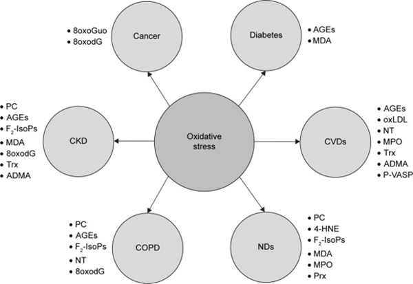
The above illustration is from the following article: Liguori, Ilaria et al. “Oxidative stress, aging, and diseases.” Clinical interventions in aging vol. 13 757-772. 26 Apr. 2018
Oxidative stress and fertility
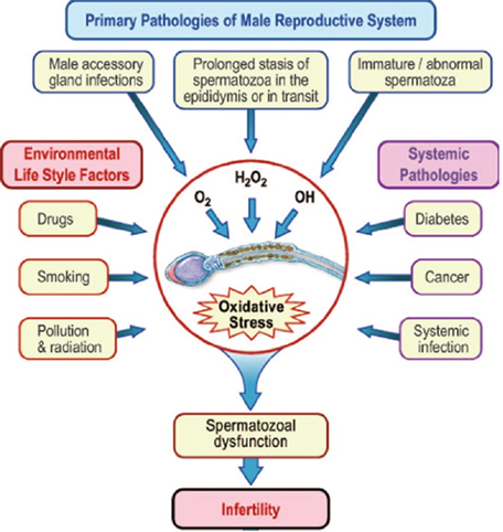
The above illustration is from the following article: Agarwal, Ashok, and Ahmad Majzoub. “Laboratory tests for oxidative stress.” Indian journal of urology : IJU : journal of the Urological Society of India vol. 33,3 (2017): 199-206.
Oxidative stress is the primary cause of male infertility. Spermatozoa are especially sensitive to exogenous toxins and external stressors. They are also able to produce their own reactive species, contributing to an unbearable oxidative burden. Direct and indirect assays can help assess seminal oxidative damage. Sensors can measure oxidation-reduction/redox potential along with additional laboratory assay including[6]
- Myeloperoxidase test
- Lipid peroxidation test
- Oxidation-reduction potential
- Chemokines
- Total antioxidant potential
Considering a combination of biomarkers along with a full assessment of oxidative stress risk factors provides the most comprehensive functional assessment of oxidative stress.
Different strategies to assess in vivo oxidative stress
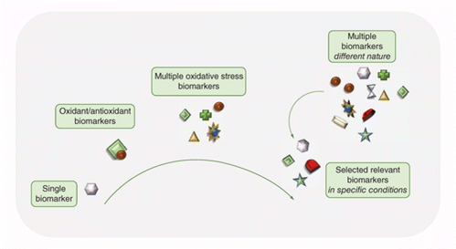
The above illustration is from the following article: Gaggini, Melania et al. “Conventional and innovative methods to assess oxidative stress biomarkers in the clinical cardiovascular setting.” BioTechniques vol. 68,4 (2020): 223-231.
|
Pros and cons of common oxidative methods. |
||
Oxidative method |
Pros |
Cons |
|
MDA |
• Availability of different methods |
• Variability in sensitivity/specificity, according to methods |
|
F2-IsoP |
• Availability of different methods |
• Ex vivo production |
|
d-ROM |
• Ease of performance |
• Ex vivo production |
|
TEAC |
• Applicability for water- or lipid-soluble antioxidants |
• Reduced power compared with real antioxidant capacity estimation |
|
ORAC |
• Ease of performance |
• Fluorescence instrumentation required |
|
FRAP |
• Reliability for: |
• Minor efficacy with albumin and glutathione |
|
Oxy-adsorbent |
• Reliability for: |
• Lack of concordance with other antioxidant assays |
LEGEND
CV: Cardiovascular; d-ROM: Diacron reactive oxygen metabolite; F2-IsoP: F2-isoprostane; FRAP: Ferric ion reducing antioxidant power assay; GC-MS: Gas chromatography/tandem mass spectrometry; MDA: Malondialdehyde; ORAC: Oxygen radical absorbance capacity assay; TEAC: Trolox equivalent antioxidant capacity assay.
The above table of information is from the following article: Gaggini, Melania et al. “Conventional and innovative methods to assess oxidative stress biomarkers in the clinical cardiovascular setting.” BioTechniques vol. 68,4 (2020): 223-231.
The use of specialized markers of oxidative stress will have limitations, including availability and specificity and should always be assessed within a larger clinical picture. Clusters of biomarkers and related patterns are more helpful than single biomarker results.
Researchers suggest “as many of the markers have been measured in similar diseases, a combination of them in large-scale panels and pattern analysis could provide an additional approach to measure disease progression or therapeutic outcome… Measurement of larger panels of biomarkers in key conditions will help give a more comprehensive picture of their significance.”[7]
Although there is still a lack of consensus regarding standardization of measuring specific markers of oxidative stress,[8] [9] some commercial labs provide assessment of related markers including[10] [11]
- Cysteine
- Cystine
- Glutathione
- Glutathione, Peroxidase
- Lipid Peroxides
- Sulfate
- Superoxide dismutase (SOD)
- Total Antioxidant Capacity, TAC
- Antioxidant assays
- DNA/RNA Damage and Repair
- Lipid peroxidation, additional
- Oxidase/peroxidase assays
- Protein oxidation/nitration
- Reactive Oxygen Species (ROS) assays
Up Next - Oxidative Stress and Cardiomatabolic Disease
References
[1] Katerji, Meghri et al. “Approaches and Methods to Measure Oxidative Stress in Clinical Samples: Research Applications in the Cancer Field.” Oxidative medicine and cellular longevity vol. 2019 1279250. 12 Mar. 2019
[2] Jain, Kewal K., and Kewal K. Jain. The handbook of biomarkers. New York: Springer, 2010.
[3] Jain, Kewal K. The handbook of biomarkers. Humana Press, New York, NY, 2017.
[4] Liguori, Ilaria et al. “Oxidative stress, aging, and diseases.” Clinical interventions in aging vol. 13 757-772. 26 Apr. 2018,
[5] Zhang, Bing, and James L Zehnder. “Oxidative stress and immune thrombocytopenia.” Seminars in hematology vol. 50,3 (2013): e1-4.
[6] Agarwal, Ashok, and Ahmad Majzoub. “Laboratory tests for oxidative stress.” Indian journal of urology : IJU : journal of the Urological Society of India vol. 33,3 (2017): 199-206.
[7] Frijhoff, Jeroen et al. “Clinical Relevance of Biomarkers of Oxidative Stress.” Antioxidants & redox signaling vol. 23,14 (2015): 1144-70.
[8] Marrocco, Ilaria et al. “Measurement and Clinical Significance of Biomarkers of Oxidative Stress in Humans.” Oxidative medicine and cellular longevity vol. 2017 (2017): 6501046.
[9] Palmieri, Beniamino, and Valeriana Sblendorio. “Current status of measuring oxidative stress.” Methods in molecular biology (Clifton, N.J.) vol. 594 (2010): 3-17.
[10] Genova Diagnostics
[11] Cell BioLabs Inc. Oxidative & Cellular Stress.


