


No biomarker is an island unto itself. Instead, it is just the tip of the metabolic iceberg…or perhaps the volcano. Ongoing research confirms the inherent value of monitoring key biomarkers and groups of biomarkers to assess health and disease…and even predict all-cause mortality.[i]
For example, elevated “prediabetes” levels of blood glucose have an impact beyond diabetes risk, and are associated with abdominal/visceral obesity, hypertension, and dyslipidemia such as high triglycerides and/or low HDL cholesterol.[ii] The takeaway can no longer be “Well, at least you don’t have diabetes.”
Endothelial dysfunction is an example of the interrelatedness of biomarkers. A common pattern associated with this disorder, which underlies atherosclerosis and most cardiovascular disease, includes:
|
Elevated |
Decreased |
|
Homocysteine[iii] [iv] Fibrinogen[v] CRP[vi] Serum iron[vii] Ferritin[viii] GGT[ix] Neutrophil/Lymphocyte ratio[x] Oxidized LDL[xi] [xii] [xiii] ADMA[xiv] Myeloperoxidase[xv] Malondialdehyde[xvi] Post-prandial glucose[xvii] Two-hour glucose in OGTT[xviii] Impaired fasting glucose and HbA1C[xix] *Oscillating glucose[xx] |
Omega-3 index[xxi] Adiponectin[xxii] Testosterone[xxiii] |
|
*Oscillating glucose between 90 and 270 mg/dL (5 and 15 mmol/L) (endothelial dysfunction was reversed by vitamin C infusion 3mg/min). |
|
Assessing key biomarkers within a matrix creates an important tool for assessing overall health, disease, and risk for all-cause and specific causes of mortality.
Readily available serum biomarkers provide a snapshot of metabolic function and include comprehensive metabolic panel, CBC with differential, lipid panel, mineral panel, hemoglobin A1c, and inflammatory biomarkers.
Researchers have begun to develop various tools to assess groups of biomarkers and their relation to disease risk and overall mortality. These include the Intermountain Risk Score (IMRS) and the Health Status Metric (HSM). The latter is found to predict all-cause mortality and specific causes of mortality such as diabetes, kidney disease, and liver disease.[xxiv]
|
Dysfunction |
Elevated |
Decreased |
|
Adrenal hyperfunction |
Sodium, chloride, CO2, BUN |
Potassium, cholesterol, triglycerides |
|
Adrenal hypofunction |
Potassium, cholesterol, triglycerides |
Sodium, chloride, blood glucose |
|
Anemia- B12/folate deficiency |
MCH, MCV, RDW, serum iron, LDH, MMA, homocysteine |
RBCs, Hct, Hgb, WBCs, neutrophils, uric acid |
|
Arthritis |
ESR, CRP, globulin, platelets, albumin (or normal) |
Decreased or normal albumin |
|
Atherosclerosis |
Increased triglycerides, cholesterol (or normal), oxidized cholesterol, small dense LDL particles, uric acid, platelets. CRP, ApoB/Apo A-1 ratio, Lp(a) |
HDL cholesterol Antioxidant status |
|
Insulin resistance |
Glucose, insulin, C-peptide, HgbA1C, total cholesterol, triglycerides, triglyceride/HDL ratio, HOMA2-IR greater than 1.8 |
Adiponectin |
|
Metabolic syndrome |
Glucose, insulin, HgbA1C, total cholesterol, triglycerides, also obesity and blood pressure |
HDL cholesterol |
|
Polycythemia |
RBCs, Hct, Hgb, total bilirubin, uric acid, WBCs, basophils, alkaline phosphatase |
MCV (or normal), MCH (or normal), serum iron (or normal) |
Reaching beyond reference intervals that define disease, it is the functional practitioner’s goal to assess these same serum biomarkers in a matrix based on optimal ranges. This approach is believed to best provide a picture of health and optimal metabolic function instead of just diagnosing disease and predicting mortality. Such an assessment provides the clinician with a valuable tool to detect subtle preclinical changes that can then be addressed and corrected.
Researchers note that ranges and patterns are not diagnostic in, and of themselves, and must be combined with other aspects of a patient’s clinical picture. However, it is equally important to recognize that many chronic diseases share similar biochemical patterns that should be fully investigated and addressed in order to optimize physiological function.
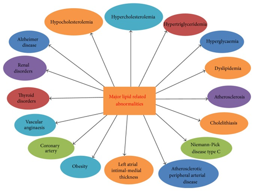
Source: Upadhyay, Ravi Kant. “Emerging risk biomarkers in cardiovascular diseases and disorders.” Journal of lipids vol. 2015 (2015): 971453. doi:10.1155/2015/971453 This is an open access article distributed under the Creative Commons Attribution License, which permits unrestricted use, distribution, and reproduction in any medium, provided the original work is properly cited.
[i] Peto, Maximus V et al. “MortalityPredictors.org: a manually-curated database of published biomarkers of human all-cause mortality.” Aging vol. 9,8 (2017): 1916-1925. doi:10.18632/aging.101280 Unrestricted Creative Commons license
[ii] American Diabetes Association. “2. Classification and Diagnosis of Diabetes: Standards of Medical Care in Diabetes-2020.” Diabetes care vol. 43,Suppl 1 (2020): S14-S31. doi:10.2337/dc20-S002
[iii] Bendall, Jennifer K et al. “Tetrahydrobiopterin in cardiovascular health and disease.” Antioxidants & redox signaling vol. 20,18 (2014): 3040-77. doi:10.1089/ars.2013.5566
[iv] University of Michigan. Pathology Handbook. Accessed October 25, 2020 from
[v] Ellins, Elizabeth A et al. “Increased fibrinogen responses to psychophysiological stress predict future endothelial dysfunction implications for cardiovascular disease?.” Brain, behavior, and immunity vol. 60 (2017): 233-239. doi:10.1016/j.bbi.2016.10.017
[vi] Anderson, Todd J. “Nitric oxide, atherosclerosis and the clinical relevance of endothelial dysfunction.” Heart failure reviews vol. 8,1 (2003): 71-86. doi:10.1023/a:1022199021949
[vii] Duffy, S J et al. “Iron chelation improves endothelial function in patients with coronary artery disease.” Circulation vol. 103,23 (2001): 2799-804. doi:10.1161/01.cir.103.23.2799
[viii] Sciacqua, Angela, et al. "Effect modification by ferritin on the relationship between inflammation and arterial stiffness in hypertensive patients with different glucose tolerance." (2020).
[ix] Bradley, Ryan D et al. “Associations between γ-glutamyltransferase (GGT) and biomarkers of atherosclerosis: the Multi-ethnic Study of Atherosclerosis (MESA).” Atherosclerosis vol. 233,2 (2014): 387-393. doi:10.1016/j.atherosclerosis.2014.01.010
[x] Martínez-Urbistondo, Diego et al. “The neutrophil-to-lymphocyte ratio as a marker of systemic endothelial dysfunction in asymptomatic subjects.” “El índice neutrófilo/linfocito como marcador de disfunción sistémica endotelial en sujetos asintomáticos.” Nefrologia : publicacion oficial de la Sociedad Espanola Nefrologia vol. 36,4 (2016): 397-403. doi:10.1016/j.nefro.2015.10.018
[xi] Chen, Chong, and Damir B Khismatullin. “Oxidized low-density lipoprotein contributes to atherogenesis via co-activation of macrophages and mast cells.” PloS one vol. 10,3 e0123088. 26 Mar. 2015, doi:10.1371/journal.pone.0123088
[xii] Gao, Shen et al. “Circulating Oxidized Low-Density Lipoprotein Levels Independently Predict 10-Year Progression of Subclinical Carotid Atherosclerosis: A Community-Based Cohort Study.” Journal of atherosclerosis and thrombosis vol. 25,10 (2018): 1032-1043. doi:10.5551/jat.43299
[xiii] Chen, Chong, and Damir B Khismatullin. “Oxidized low-density lipoprotein contributes to atherogenesis via co-activation of macrophages and mast cells.” PloS one vol. 10,3 e0123088. 26 Mar. 2015, doi:10.1371/journal.pone.0123088
[xiv] Widmer, R Jay, and Amir Lerman. “Endothelial dysfunction and cardiovascular disease.” Global cardiology science & practice vol. 2014,3 291-308. 16 Oct. 2014, doi:10.5339/gcsp.2014.43 This is an open access article distributed under the terms of the Creative Commons Attribution license CC BY 4.0, which permits unrestricted use, distribution and reproduction in any medium, provided the original work is properly cited.
[xv] Upadhyay, Ravi Kant. “Emerging risk biomarkers in cardiovascular diseases and disorders.” Journal of lipids vol. 2015 (2015): 971453. doi:10.1155/2015/971453 This is an open access article distributed under the Creative Commons Attribution License, which permits unrestricted use, distribution, and reproduction in any medium, provided the original work is properly cited.
[xvi] Yang, Rui-Li et al. “Increasing Oxidative Stress with Progressive Hyperlipidemia in Human: Relation between Malondialdehyde and Atherogenic Index.” Journal of clinical biochemistry and nutrition vol. 43,3 (2008): 154-8. doi:10.3164/jcbn.2008044
[xvii] Whisner, Corrie M et al. “Effects of Low-Fat and High-Fat Meals, with and without Dietary Fiber, on Postprandial Endothelial Function, Triglyceridemia, and Glycemia in Adolescents.” Nutrients vol. 11,11 2626. 2 Nov. 2019, doi:10.3390/nu11112626
[xviii] Tomiyama, H et al. “Close relationship of abnormal glucose tolerance with endothelial dysfunction in hypertension.” Hypertension (Dallas, Tex. : 1979) vol. 36,2 (2000): 245-9. doi:10.1161/01.hyp.36.2.245
[xix] Vehkavaara, S et al. “In vivo endothelial dysfunction characterizes patients with impaired fasting glucose.” Diabetes care vol. 22,12 (1999): 2055-60. doi:10.2337/diacare.22.12.2055
[xx] Ceriello, Antonio et al. “Oscillating glucose is more deleterious to endothelial function and oxidative stress than mean glucose in normal and type 2 diabetic patients.” Diabetes vol. 57,5 (2008): 1349-54. doi:10.2337/db08-0063
[xxi] Zehr, Kayla R, and Mary K Walker. “Omega-3 polyunsaturated fatty acids improve endothelial function in humans at risk for atherosclerosis: A review.” Prostaglandins & other lipid mediators vol. 134 (2018): 131-140. doi:10.1016/j.prostaglandins.2017.07.005
[xxii] Hui, Xiaoyan et al. “Adiponectin and cardiovascular health: an update.” British journal of pharmacology vol. 165,3 (2012): 574-90. doi:10.1111/j.1476-5381.2011.01395.x
[xxiii] Akishita, Masahiro et al. “Low testosterone level is an independent determinant of endothelial dysfunction in men.” Hypertension research : official journal of the Japanese Society of Hypertension vol. 30,11 (2007): 1029-34. doi:10.1291/hypres.30.1029
[xxiv] Bello, Ghalib A et al. “Development and Validation of a Clinical Risk-Assessment Tool Predictive of All-Cause Mortality.” Bioinformatics and biology insights vol. 9,Suppl 3 1-10. 1 Sep. 2015, doi:10.4137/BBI.S30172


